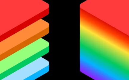
Every object or substance in the universe reflects light slightly differently, making it possible to learn information about it. The human eye and traditional cameras dismiss much of this information by condensing it down into three channels, red, green, and blue. While this lets us see colours, it is possible to see a lot more by looking a little closer.
The intricate dance between light and the structure of an object provides considerable spectral information, allowing us to uncover many properties beyond just colour.
Hyperspectral Imaging (HSI) ramps up the resolution to observe light in much more detail using 30+ channels rather than just three. With this level of detail, it is possible to detect spectral features and signatures that open up a range of new use cases, and one of the most active fields is healthcare.
Hyperspectral imaging in healthcare
HSI can detect both spatial (size and shape) and spectral features of tissue without introducing additional substances (e.g., fluorescent dyes), using ionising radiation (e.g., X-ray, CT, PET) or large equipment (e.g., MRI). These features provide data useful for both diagnostics and treatment.
Light can interact with tissue via a number of different mechanisms, including absorption, scattering, and transmission. Organic molecules often have distinct absorption and scattering properties correlated to their biochemical features.
Therefore, specific tissue properties are observable using hyperspectral systems to analyse their spectral response to light. For example, HSI can provide biological information related to:
- Attenuation from haemoglobin (one of the primary attenuators in tissue) to detect blood vessels and quantify blood oxygen levels.
- The reradiation of longer wavelength light from specific biomolecules (known as autofluorescence) that changes during disease progression.
- How tissue morphology changes during disease progression and the impact this has on its scattering properties, generating diagnostic markers.
So what would HSI in healthcare look like? The technology’s use cases can be divided into two main categories: diagnostics and surgery.
Diagnostic use cases
Diseases and injuries create a biological response in the body. In certain scenarios, these responses lead to a measurable change in the optical properties of tissue. HSI offers enough detail (i.e., significantly more data points per pixel) to accurately detect these changes and aid the diagnostic process. Listed below are three diagnostic examples of HSI in healthcare showing significant promise.
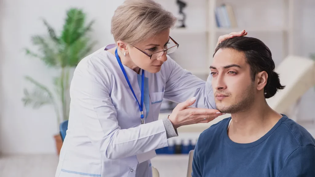
Skin cancer triage
Multi-spectral imaging systems have previously been utilized for dermoscopic assessments. However, these used fewer bands than HSI cameras and struggled with low specificity (true negative rates), overestimating the number of cancerous lesions.
Dermoscopy with trained professionals remains the best method for detecting and diagnosing melanoma and other skin cancers. But achieving high sensitivity (true positive rate) and high specificity requires dermatologists with significant clinical experience.
Given it is unlikely most of the general population has access to highly experienced dermatologists, an opportunity remains for a technological solution to improve the triage of potential skin cancer patients, reducing false positives (increasing sensitivity).
Combining HSI data with machine learning analysis could offer a non-invasive solution for triaging skin cancer patients, recommending patients of concern for biopsy and further assessment by an experienced dermatologist. This type of solution could increase sensitivity, ensuring medical practitioners make the most of their time, focusing on the patients who truly need their help.
In recent years a number of preliminary and proof of concept studies have investigated the potential for HSI in skin cancer diagnosis with promising results. This includes:
- A sensitivity of 87.5% and specificity of 100% distinguishing between malignant and benign pigmented skin lesions.
- A sensitivity of 95% and specificity of 92% training a neural network to distinguish between histopathologically verified malignant and benign melanocytic lesions based on HSI data.
Assessing burns
Assessing the severity of a burn wound is a challenging clinical process. The depth of a burn (how many layers of the skin are damaged) determines the treatment the patient receives. First-degree burns only damage the upper layers (epidermis) and can be left to heal with supportive care alone. In contrast, third-degree burns that damage the epidermis and full dermis require surgical management.
While first and third-degree burns are relatively easy to diagnose, second-degree burns (damaging the epidermis and part of the dermis) present diagnostic and treatment challenges. Some require surgical management while others don’t, and even experienced burn surgeons can struggle to distinguish between the two. Previous research suggests only 50-75% of these burns are correctly differentiated, leading to sub-optimal treatment.
As well as offering higher spectral resolution, HSI extends the light range that can be imaged beyond the visible spectrum. HSI with near-infrared (NIR) light (that penetrates deeper into the skin, unlike visible light) could offer healthcare professionals the data they need to distinguish between superficial partial burns and deep partial burns, improving patient treatment.
A paper from 2022 shows HSI could be a useful tool for burn depth assessment. The researchers compared burn patients where secondary wound closure was complete within 21 days to those who required longer or needed skin grafting. HSI was performed on patients 24 hours, 48 hours, and 72 hours after their injuries. The data generated showed that changes in microcirculation in the first 72 hours, detected using HSI, could predict the need for skin grafting with a sensitivity of 92% and a specificity of 71%.
Early-stage detection of Alzheimer’s disease
There are no reliable ways to diagnose Alzheimer’s disease (AD) (the most common cause of dementia) in its early stages. Catching it early, before the onset of cognitive or behavioural deficits, is critical to the future development of effective treatments.
Researchers know the pathogenesis of AD is caused by the build-up of specific amyloid β-protein plaques and tau neurofibrillary tangles in the brain tissue. This build-up can begin 15-20 years before the emergence of symptoms. As a result, many studies are trying to identify minimally invasive biomarkers that will aid early-stage detection.
A significant area of interest is imaging the retina using a hyperspectral camera to try and detect any changes caused by amyloid β-protein plaque deposition.
As the eyes are constantly moving, this requires a specific type of HSI called snapshot, simultaneously measuring both spectral and spatial information. The more common scanning forms of HSI cannot gather both forms of information in a single measurement, and any eye movement will ruin the final image.
Surgical use cases
The spectral information provided by HSI, beyond simple colour data of traditional systems, leads to potential use cases during surgery.
Precision surgery
Using HSI intraoperatively has the potential to improve surgical precision. Real-time hyperspectral systems can provide spectral information to help surgeons visualise different organs and structures, leading to better outcomes and avoiding complications. For example, differentiating between ureter and adipose tissue during colorectal surgery.
Tumour resection
Removing tumours surgically, a process called resection is a crucial part of cancer management for many patients. A significant predictive factor as to the patient’s survival post-surgery is the completeness of the tumour’s removal, i.e., the removal of all tumour cells.
This means the surgeon has to be able to accurately identify the edges of a tumour during the operation. A difficult challenge, surgeons generally rely on visual appearance and manual palpation when differentiating between healthy and tumour cells.
Reports show HSI offers a real-time solution to identifying tumour margins during surgery. HSI could improve the outcome of future resection with the doctor able to precisely remove all cancer cells without harming healthy tissue.
Monitoring
During surgery, the patient’s blood flow has to be monitored (a process called ischemia) to prevent tissue damage or more significant complications that may lead to death. HSI systems have the potential to deliver real-time intraoperative blood flow monitoring. This relies on HSI’s ability to determine tissue oxygenation.

Progress of HSI in healthcare
While use of HSI techniques in health care dates back to the early 2000s, recent technological advancements have dramatically increased interest. At Living Optics, we are looking to advance HSI to deliver the performance and reliability needed for multiple healthcare use cases.
To learn more about our next-generation snapshot hyperspectral system, contact us to schedule a demo .